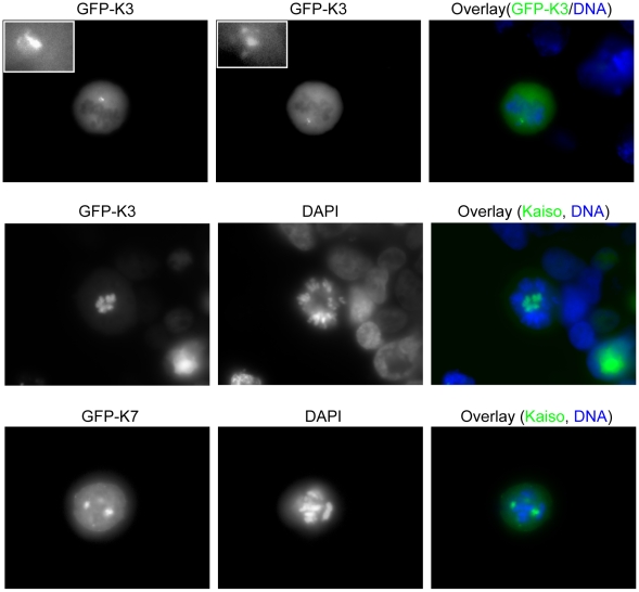
Figure 9. The GFP-tagged Kaiso fragments K3 and K7 abnormally localize at the centrosomes and cause aberrant chromosomal distribution.
HEK293 cells were transfected with constructs GFP-K3 and GFP-K7 (see also Fig. 1). In the upper row, cells were imaged by focusing on, respectively, the upper centrosomal region (left) and the lower centrosomal region (middle) (see also insets for magnification of these regions); an overlay of the upper middle picture with DAPI staining to detect DNA is shown on the right. Pictures in the lower two rows also reveal abnormal chromosomal distributions upon overexpression of, respectively, Kaiso fragments K3 and K7. DNA was stained with DAPI. A Zeiss Axiophot microscope was used (100× objective lens).

