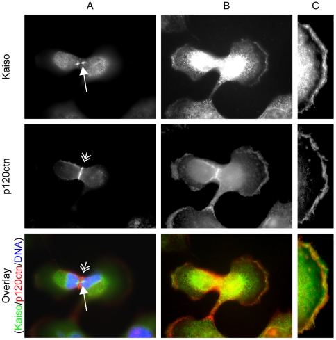
Figure 10. Kaiso and p120ctn localization during cytokinesis.
Column A shows pictures made after focusing on the midbody of SK-LMS-1 cells: Kaiso is present at the midbody (long arrow) while p120ctn is located at the cleavage furrow (double short arrow) between the two forming cells. Column B shows pictures of the same cells while focusing on the protrusions at the cell borders. Kaiso and p120ctn partly colocalize there. Pictures in column C are magnifications of the free edge of one of the daughter cells. Cells were stained with a polyclonal antibody (S1337) for Kaiso detection, and with a monoclonal antibody (pp120) for p120ctn detection, followed by an Alexa 488-conjugated anti-rabbit secondary antibody for Kaiso, and an Alexa 594-conjugated anti-mouse secondary antibody for p120ctn. DNA was stained with DAPI. Cells were imaged (63× objective lens) with a Zeiss Axiophot microscope.

