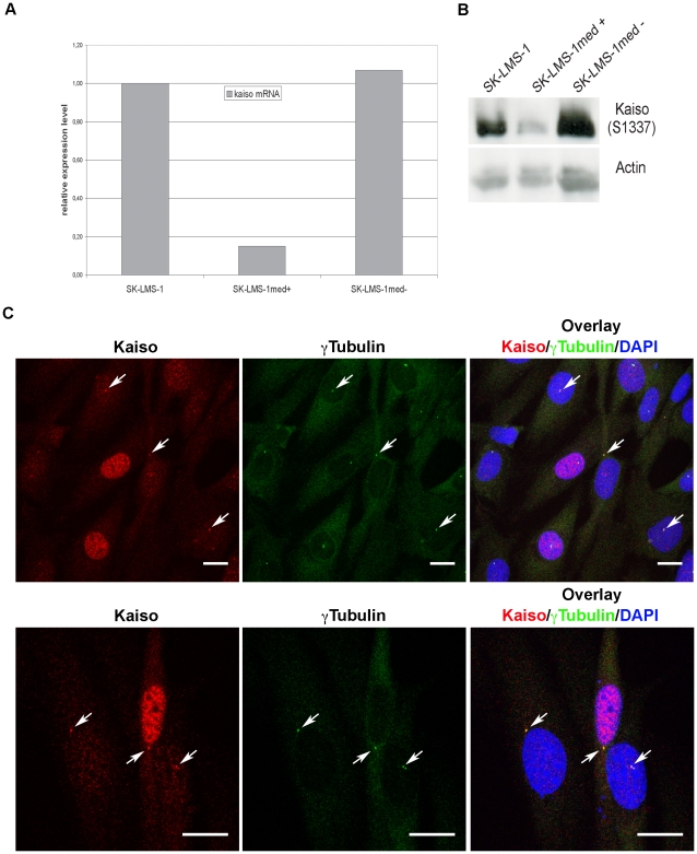
Figure 11. Knockdown of Kaiso in SK-LMS-1_shKAISO cells affects mainly nuclear Kaiso.
Parental SK-LMS-1 cells were transduced with lentiviruses encoding transcripts comprising Kaiso-specific shRNA and GFP mRNA. Cells were then FACS sorted and cell populations with medium GFP content were resorted into GFP-positive and GFP-negative cells, named, respectively, SK-LMS-1med+ and SK-LMS-1med–. (A) Relative Kaiso-mRNA expression levels as determined by QRT-PCR. (B) Expression levels of Kaiso protein as determined by western blotting using pAb S1337 and anti-actin for normalization. (C) Immunofluorescent staining for Kaiso and γ-tubulin in SK-LMS-1_shKAISO cell populations in which part of the cells show efficient Kaiso knockdown. In the latter cells diffuse nuclear staining with Kaiso-specific pAb S1337 is gone, whereas staining in centrosomal regions is largely preserved (arrows). Similar results were obtained when staining with mAb 6F. Note that upon methanol treatment the GFP signal disappears from the cells by diffusion. DNA was stained with DAPI and cells were imaged with a Leica SP5 confocal microscope. Each figure is a projection of a confocal image stack. Scale bar, 20 µm.

