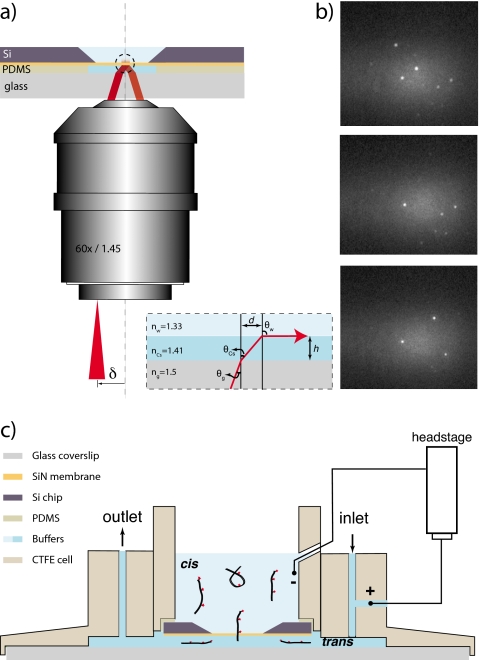
Figure 1.
(a) Schematic illustration (not to scale) of the TIR method for illumination of a thin solid membrane (orange) immersed between two aqueous fluids (light blue). Corresponding indices of refraction are indicated as nCs and nW. The system is designed such that TIR occurs at the SiN surface, enabling single-molecule detection. Inset shows ray path of an arbitrary ray incident at the glass-CsCl interface. θg, θCs, and θw are the ray angles at different interfaces, and d is the shift in the center of field of view caused due to in the intermediate CsCl column of height h. (b) Images of the SiN membrane where individual DNA molecules conjugated to single ATTO647N fluorophores are immobilized. (c) A schematic illustration of the flow cell. Outer cell and the inset are made from CTFE. Thin layers of fast curing PDMS are used to glue the silicon chip and the glass coverslip to the insert and outer cell, respectively. Inlet and outlet flow channels are used to transfer fluid in the trans chamber. Ionic current through the nanopore is measured using two Ag∕AgCl electrodes immersed in the cis and trans chambers as shown.

