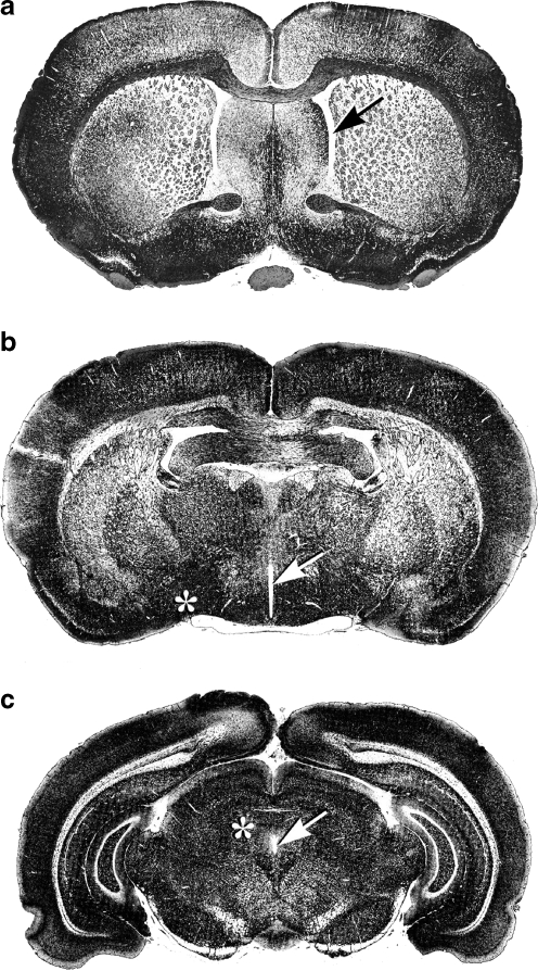
Fig. 1.
Representative frontal sections of rat brain. Frontal sections of rat brain (Sudan black staining) at three different levels from rostral a to caudal c showing: in a the lateral ventricles (arrow), in b third ventricle (arrow) and thalamus-hypothalamus with supraoptic nucleus (asterisk), and in c showing aqueduct (arrow) and periaquedutcal gray area (asterisk). ET-1 adiministration to such regions has cardiovascular (D’Amico et al. 1996; McAuley et al. 1996; Macrae et al. 1991a, b; Macrae et al. 1993) and temperature-regulating effects (Fabricio et al. 2005)

