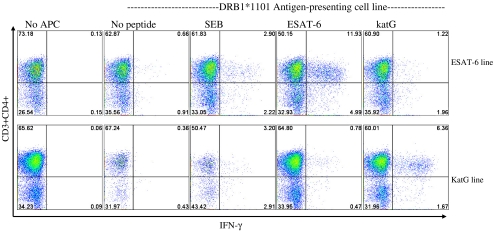
Fig. 4.
HLA-DRB1*1101 presents ESAT-6 to sarcoidosis CD4+ T cells. Intracellular cytokine staining for IFN-γ was performed on expanded ESAT-6 and katG cell lines derived from sarcoidosis subject 6, after stimulation with either ESAT-6 peptide 14, katG peptide 13, or SEB. No recognition was observed in the expanded cell lines alone, demonstrating the absence of baseline IFN-γ production. Expanded cells stimulated with ESAT-6 or katG peptide in the absence of Sweig cells revealed minimal responses, confirming the loss of antigen-presenting cells during expansion. Significant responses to ESAT-6 and katG were observed in the respective cell line when antigen was presented using the DRB1*1101 expressing Sweig cell lines. Shown are representative flow cytometry dot plots indicating percentage of sarcoidosis CD4+ T cell responses to the different stimulation conditions

