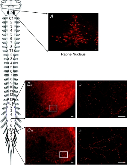
Figure 5. Serotonin positive fibres are present through out the length of the spinal cord.
A, 5-HTir positive cells in the region of the raphe nucleus were used as a positive control. B, 5-HTir positive fibres within the lumbar ventral horn (transverse slice). Ba, low power view of the ventral horn showing high level of 5-HTir expression. Bb, increased magnification of ventral horn in Ba showing detail of individual 5-HTir positive fibres and varicosities. C, 5-HTir positive fibres were also present in the sacral ventral horn. Ca, low power view of the ventral horn. Cb, increased magnification of boxed area in Ca showing individual 5-HTir fibres and varicosities. Scale bar = 20 μm. Schematic diagram shows lumbar and sacral segments selected.

