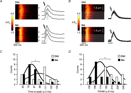
Figure 4. Increased spatial synchronization of whole-cell SR Ca2+ release in Iso is revealed at near-threshold triggers.
A, near-threshold SR Ca2+ release kinetics exhibited clear spatial inhomogeneities in control, as indicated by the temporal Ca2+ transient profiles from 3 different (subcellular) regions. Iso synchronized SR Ca2+ release throughout the cell, as reflected by the coordinated peaks of the corresponding subcellular Ca2+ transient profiles. B, the line-scan images in control and Iso in A were divided into 20 equal parts (each 1.8 μm wide), on which a detailed analysis of the temporal characteristics was performed. C, the distribution of the time to peak in the subcellular regions revealed a significantly shorter average time to peak in Iso (99.3 ± 5.4 ms in control to 78.9 ± 3.0 ms in Iso), and was less spread in time throughout the cell (reflected by the relative widths and amplitudes of the representative, integral-normalized Gaussians). D, accelerated release and decay kinetics in Iso are also reflected in shorter average full duration at half-maximum (FDHM) amplitude throughout the cell (207.6 ± 6.3 ms in control to 168.0 ± 5.0 ms in Iso) (*P < 0.01).

