Figure 7.
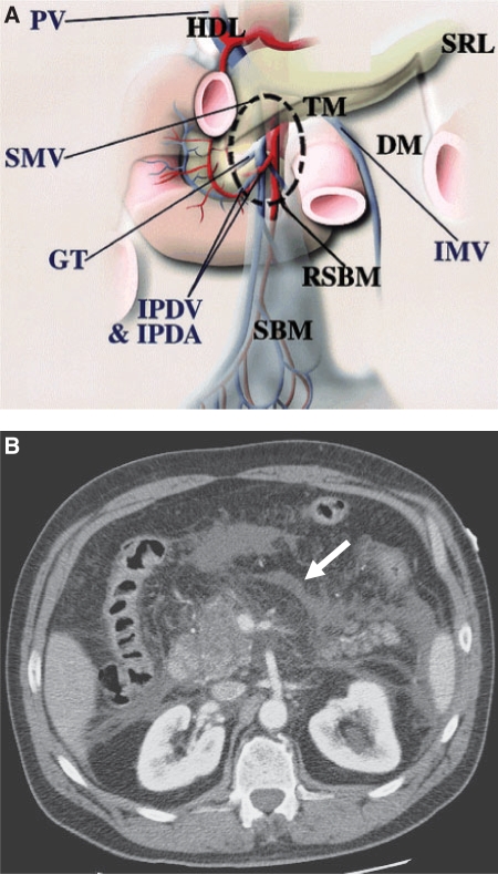
Small bowel mesentery: conduit of disease. (A) The anatomy near the root of the SBM (RSBM). The root of the SBM (area within dashed circle) is contiguous superiorly to the hepatoduodenal ligament (HDL) along the SMV, anteriorly to the transverse mesocolon (TM), and posterolaterally to the ascending mesocolon and descending mesocolon (DM). The gastrocolic trunk (GT) is a landmark of the junction between the transverse mesocolon and the root of the SBM. The inferior mesenteric vein (IMV) is a landmark of the descending mesocolon and joins the SMV or splenic vein on the left side of the root of the SBM. IPDA, inferior pancreaticoduodenal artery; IPDV, inferior pancreaticoduodenal vein; PV, portal vein; SRL, splenorenal ligament. (From Okino Y, Kiyosue H, Mori H, et al. Root of the small-bowel mesentery: correlative anatomy and CT features of pathologic conditions. Radiographics 2001; 21: 1476; with permission.) (B) CT scan shows fluid (arrow) in the subperitoneal space of the small bowel mesentery in this patient with pancreatitis.

