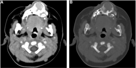
Figure 2.
Axial contrast-enhanced CT scan images in (A) soft-tissue window and (B) bone window settings show an expansile, bilobed, well-circumscribed, solid lesion with epicenter in the anterior maxillary alveolar ridge. Bone margins are continuous, thin, lobulated with areas of sclerosis and hyperostosis. Moderate soft-tissue enhancement is present. Note the displaced developing teeth.

