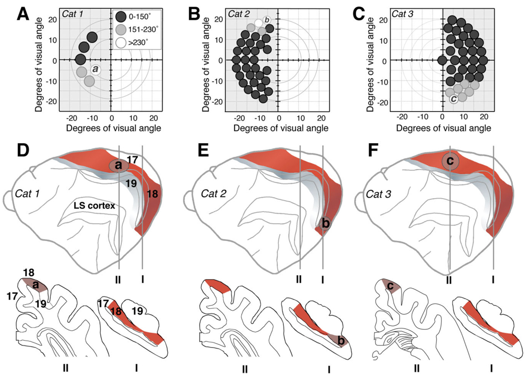
Fig. 4.
Retinotopy of training-induced visual recovery in Cats 1, 2, and 3. A–C: Maps of the visual field in Cats 1, 2, and 3 illustrating, via the indicated gray scale, the magnitude of training-induced improvements in direction range thresholds (relative to immediate postlesion performance) at locations “a”, “b,” and “c” within the contralesional, impaired hemifields, as well as at several adjacent locations. Circles (drawn to scale) represent the size and position of random dot stimuli used to measure direction range thresholds at each of the locations tested. White circles denote the largest improvements attained, which averaged above 230° of direction range and resulted in recovery to normal levels of performance relative to equivalent locations in the intact hemifield. Light gray circles indicate moderate improvements to less than normal levels of performance. Dark gray circles denote little to no improvement. D–F: Schematic diagrams indicating the approximate locations of regions within area 18 that corresponded retinotopically to retrained locations “a,” “b,” and “c” in Cats 1 (D), 2 (E), and 3 (F), respectively. The lateral views of the cat brain are displayed in an “open sulcus” configuration, and illustrate the location of cortical area 18 (red shading) relative to areas 17 (white), 19 (gray), and LS cortex (damaged in all three cats). Coronal sections (traced by using the NeuroLucida software) from locations I (~7 mm posterior to the interaural line) and II (~1 mm anterior to the interaural line for Cat 2 and ~8 mm anterior to the interaural line for Cat 3) illustrate the different retinotropic regions of area 18 selected for analysis. According to the electrophysiological maps of Tusa and colleagues (1979, 1981), the area 18 representation of location “a” in the mid-lower field is more anterior and dorsal than the brain location of far upper field region “b,” but it is more posterior than the brain location where the far lower field location “c” is represented.

