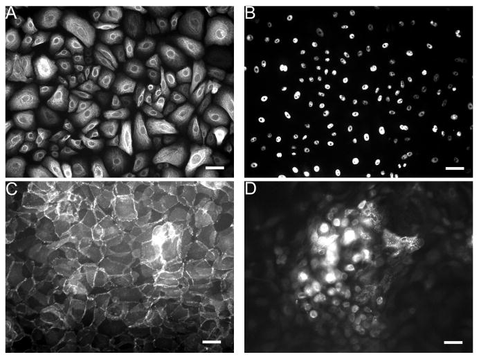
Figure 1.
Immunofluorescent staining of hES cell-derived keratinocyte progenitors. A, B) Subcultured keratinocytes (as in step 3.1.5. of protocol) immunostained against the cytoskeletal protein K14 (A) and the nuclear transcription factor p63 (B). C) Differentiated hES cells (after 3 weeks culture in step 3.1.4. of protocol) stained against β-catenin; note punctate staining at membranes. D) Keratinocyte progenitors cultured to confluence and treated with 0.8 mM Ca2+ (after 4 weeks of culture in step 3.1.4. of protocol), then stained against the terminal differentiation marker filaggrin; note localization within suprabasal layers. Scale bar denotes 50 μm.

