Figure 9.

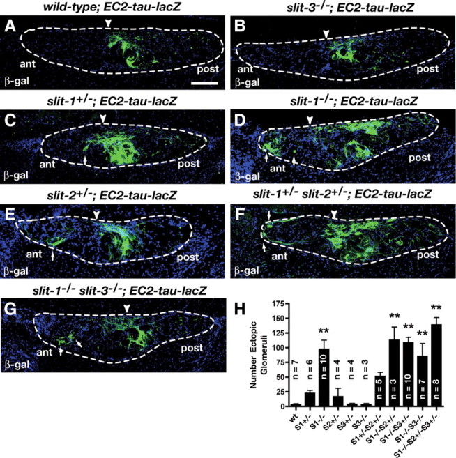
Differential contributions of Slits to the targeting of vomeronasal projections. A–G, Parasagittal AOB sections of adult wild-type; EC2-tau-lacZ (A), slit-3−/−; EC2-tau-lacZ (B), slit-1+/−; EC2-tau-lacZ (C), slit-1−/−; EC2-tau-lacZ (D), slit-2+/−; EC2-tau-lacZ (E), slit-1-+/− slit-2+/−; EC2-tau-lacZ (F), and slit-1−/− slit-3−/−; EC2-tau-lacZ (G) mice stained with anti-β-galactosidase. In wild-type mice, EC2-expressing axons innervate the posterior region of the AOB (A) (7 of 7). A subset of EC2-expressing axons are mistargeted to the anterior AOB in slit-1+/−; EC2-tau-lacZ mice (C) (3 of 6), slit-1−/−; EC2-tau-lacZ mice (D) (10 of 10), slit-2+/−; EC2-tau-lacZ mice (E) (1 of 4), slit-1+/− slit-2+/−; EC2-tau-lacZ mice (F) (5 of 5), and slit-1−/− slit-3−/−; EC2-tau-lacZ mice (G) (7 of 7). In slit-3−/−; EC2-tau-lacZ mice all EC2-expressing axons target accurately to the posterior AOB (B) (3 of 3). Dotted lines outline the nerve and glomerular layers of the AOB and arrowheads indicate the anterior–posterior border as defined by BS lectin staining (data not shown). Scale bar, 125 μm. H, Quantification of ectopic innervation of the AOB in control and slit mutant mice. Data were analyzed by a Dunnett one-way ANOVA. Double asterisk, p < 0.01 versus wild-type mice. n values are indicated on the figure.
