
Figure 3. REP 9 does not interfere with LCMV virus production.
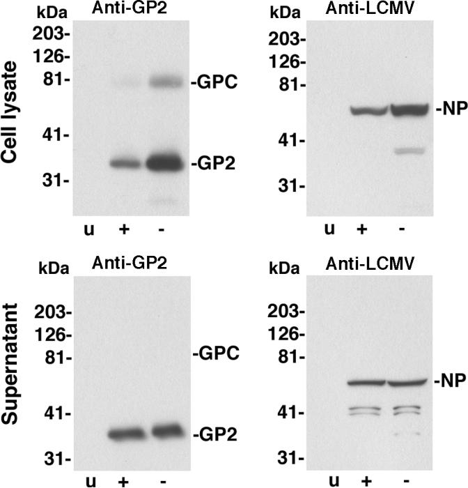
VeroE6 cells were infected with LCMV ARM53b at an MOI of 1 or mock infected (u). After 24 hours, medium was removed, cells washed extensively and new medium added with (+) or without (-) 2 μM REP 9. After 24 hours, total cell lysates were prepared. Supernatants were subjected to ultracentrifugation through a sucrose cushion and pellets solubilized in SDS-PAGE sample buffer. Total proteins of cell lysates (cell lysate) and pellets recovered from supernatants by ultracentrifugation (supernatant) were separated by SDS-PAGE, blotted to nitrocellulose and probed with mAb 83.6 to LCMVGP2 (anti-GP) and polyclonal guinea pig serum anti-LCMV. Primary antibodies were detected with HRP-conjugated secondary antibodies using enhanced chemiluminescence (ECL). Molecular masses and the positions of GPC, GP2, and NP are indicated.