Fig. 2.
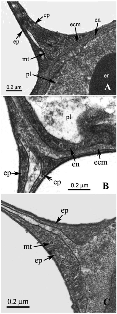
A. High-power EM of a junction of an epithelial bridge with a capillary wall. Marked enlargement of one of the epithelial cells is well shown. This section of the cell clearly shows extremely small circular inclusions that have a diameter of approximately 20 nm. These may be microtubules. The space between the two epithelial cells making up the bridge is contiguous with extracellular matrix of the capillary wall. ecm, extracellular matrix; en, endothelial cell; ep, epithelial cell; er, erythrocyte; mt, microtubule; pl, plasma. B. Another example of a junction between them an epithelial bridge and a capillary wall. Again there is some enlargement of the epithelial cells near the junction. The small circular inclusions referred to in relation to Fig. 2A are just visible. Also the space between the two epithelial cells is contiguous with extracellular matrix of the capillary wall. ecm, extracellular matrix; en, endothelial cell; ep, epithelial cell; pl, plasma. C. Another example of a junction showing the continuation of the extracellular matrix of the capillary wall into the space between the two epithelial cells of the bridge. This section was cut diagonally which explains the thickness of the extracellular matrix layer. ep, epithelial cell; mt, microtubule.

