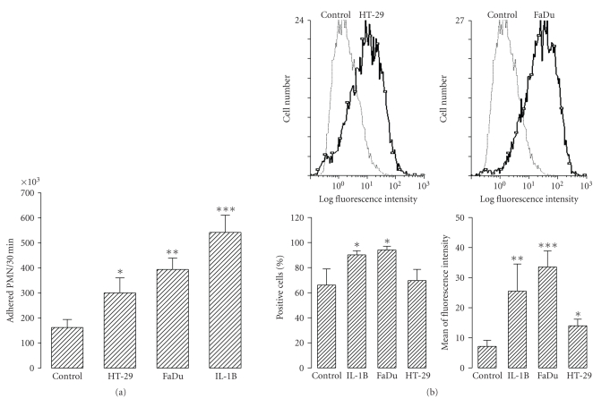
Figure 8.
(a) Adhesion of PMN to HMVEC exposed to tumour cell conditioning medium. 2 × 10⁶ DiI-labeled PMN were added to the culture wells containing confluent HMVEC previously treated overnight with none (control), HT-29 (HT-29) and FaDu (FaDu) 48 hours conditioning medium, and 10 U/mL of human recombinant IL-1β (IL-1B) and then incubated at 37°C for 30 minutes. After washing, adhered PMN were evaluated as described in the Methods section. Bars represent the mean±SEM of 7 independent experiments performed in sixtoplicate. (b) Surface expression of ICAM-1 in HMVEC exposed to tumour cell conditioning media. HMVEC previously treated overnight with none (control), HT-29 (HT-29) and FaDu (FaDu) 48 hours conditioning medium, and 10 U/mL of human recombinant IL-1β (IL-1B) were detached and analyzed for ICAM-1 expression by flow cytometry (see Methods). Upper panels represent histograms (out of 4) showing the effect of tumour cell conditioning medium on ICAM-1 expression. Bottom panels show quantitative data. Bars represent the mean ± SEM of 4 independent experiments. Statistical significance was assessed using ANOVA test; *P < .05, **P < .01, and ***P < .001 when compared to control group.

