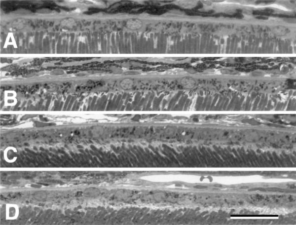
Fig. 2.
High power photomicrographs of the RPE cell layer in a wild-type mouse (A, B) and a cd81−/− mouse littermate (C, D). Notice that the RPE cell layer appears similar. The RPE sits on a well-defined basal lamina Bruch's membrane and the apical surface is intimate contact with the outer segments. All photomicrographs are taken at the same magnification and the scale bar in D = 25 μm.

