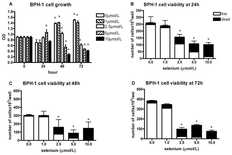
FIG. 2.
The effect of sodium selenite on BPH-1 cell growth and viability. The data represent the mean ± SD of three experiments. A: Cell growth measured by the MTT assay presented as absorbance read by microplate reader at 570 nm at different time intervals; *P < 0.001 (two-sided) for difference with control cells. OD, optical density. B–D: The number of viable cells and cell viability determined by hemocytometer counting and trypan blue exclusion after 24 h (B), 48 h (C), and 72 h (D); *P < 0.001 (two-sided) for difference with control cells.

