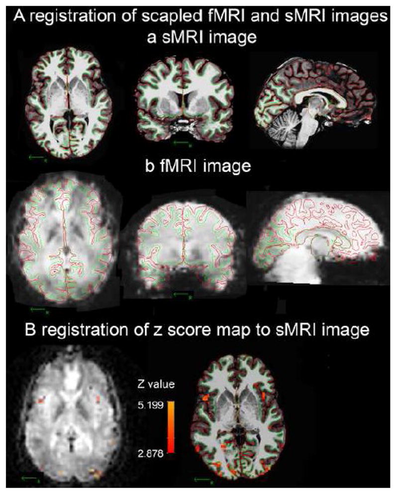
Figure 2.

Example of registration of fMRI activity onto a high quality sMRI image at the individual level. The WM/GM border (green line) and GM/pial border (red line) were created by Freesurfer in the skull-removed sMRI image. A: the borders of one subject derived from his sMRI image (a: coronal, axial and sagittal views) were registered on his fMRI template (b: coronal, axial and sagittal views). B: Registering the significant voxels of [(CS+)-fixation] from the fMRI template onto the sMRI image using final registration parameters created by procedure 4.
