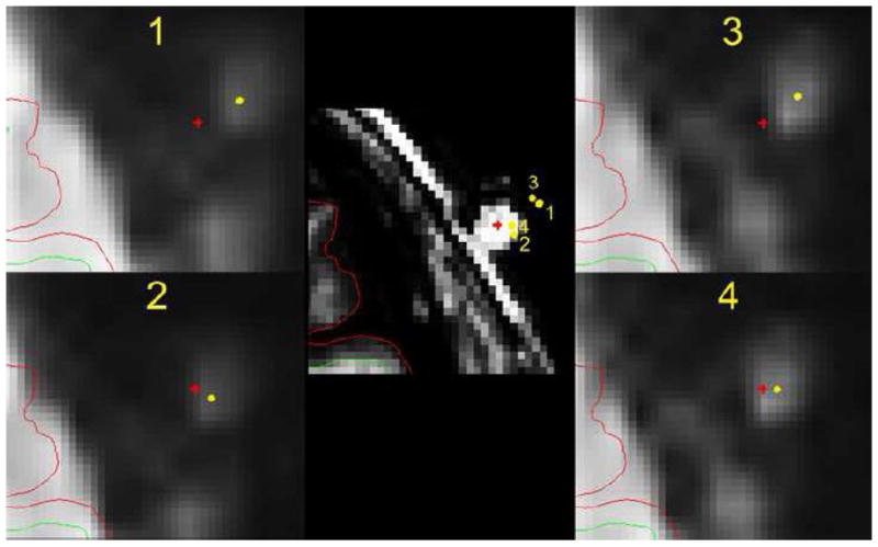
Figure 3.

Estimation of accuracy of registration procedures with an extracanonical marker. The fMRI template was registered and resampled to the sMRI with skull image using procedure 1, 2, 3, and 4 (see method). The center of the marker (the bright dot outside the brain) on this slice of the resampled fMRI template is labeled with a yellow dot. The center of the marker (the bright dot on the skull) on the same slice of sMRI image (middle) is labeled with a red cross. Procedure 1, 2, 3, and 4 produced different registration of marker center of sMRI onto fMRI slice as illustrated in each figure at periphery. The marker center of the fMRI template is registered on the sMRI slice (middle) at four locations using procedure 1, 2, 3, and 4. The diameter of the marker is about 8 mm. All other conventions are the same as in the figure 2.
