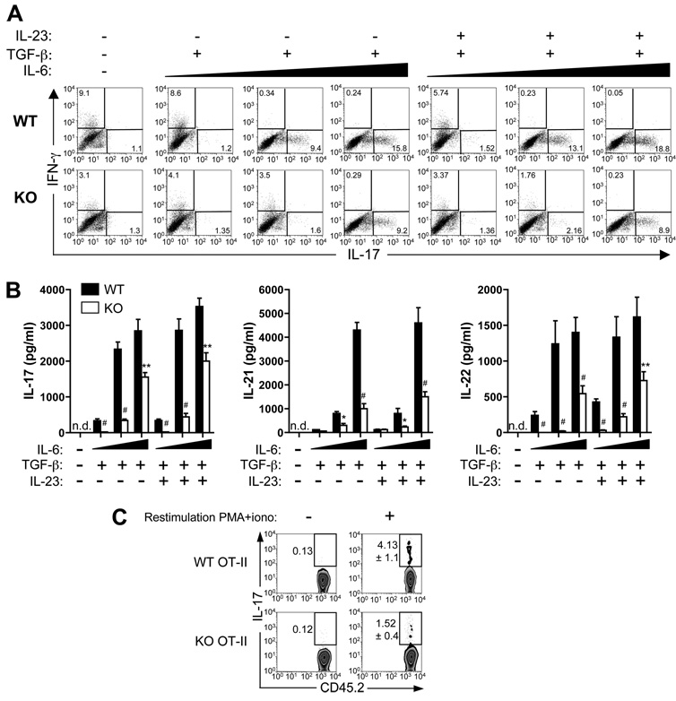
FIGURE 5.
Defective in vitro and in vivo Th17 differentiation of Def6−/− T cells. A and B, FACS-sorted naive CD4+CD62LhighCD44low OT-II T cells from WT or KO mice were stimulated for 5 days with irradiated, OVA323–339-pulsed WT splenic APCs, and cultured in the absence or presence of TGF-β (5 ng/ml) and increasing concentrations (1, 5, or 20 ng/ml; indicated by a black wedge) of IL-6. IL-17- and IFN-γ-expressing CD4+ T cells were measured by ICS (A). The production of IL-17, IL-21, and IL-22 in culture supernatants was measured by an ELISA (B). Data represent means ± SD of triplicate wells and are representative of three independent experiments with similar results. C, FACS-sorted naive CD45.2+ T cells from WT or KO OT-II mice were transferred i.v. into CD45-congenic (CD45.1+) mice. The recipients (three mice per group) were immunized with OVA in CFA. Eight days later, dLN cells were stimulated with PMA and ionomycin for 5 h, and IL-17-expressing CD4+ T cells were measured by ICS after gating on CD45.2+CD4+ T cells. Numbers adjacent to the gates indicate the percentage of IL-17-positive cells among the transferred cells. Statistical differences were determined as in Fig. 1.

