
Figure 6.
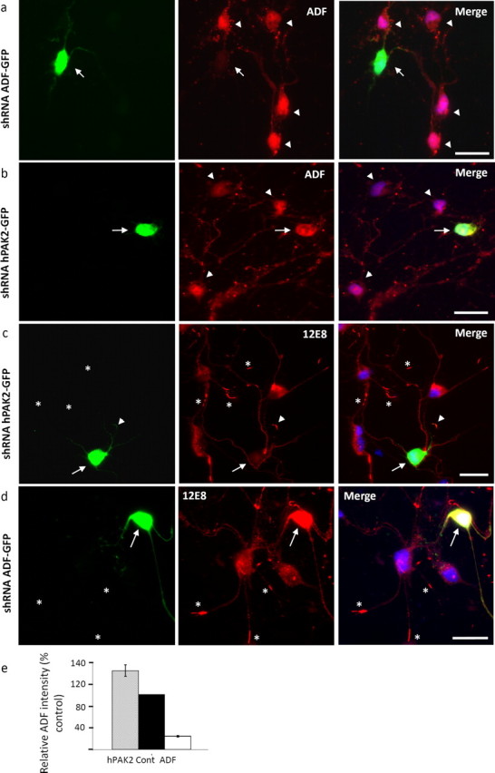
Reduction of the cellular ADF/cofilin pool inhibits the formation of rods and sequestration of pMAP. a, ADF was knocked down in primary chick tectal neurons by expression of shRNA from a plasmid also encoding GFP for visualization of transfected cells. In cultures transfected for 4 d, ADF immunolabeling using the 1439 antibody demonstrated significant ADF knockdown in transfected cells (arrow) compared with surrounding nontransfected cells (arrowheads). b, Cells transfected with an shRNA specific for human PAK2 as a control (arrow) showed no reduction of ADF compared with surrounding nontransfected cells (arrowheads). c, d, Transfected cultures were treated with 1 μm AM and immunolabeled with 12E8 to determine whether pMAP accumulates into rod-like structures after significant reduction of ADF. Whereas hPAK2 shRNA-transfected cells (c, arrow) formed pMAP-positive rods (arrowhead) comparable with surrounding nontransfected cells (asterisks), ADF shRNA-transfected cells (d, arrow) never accumulated pMAP-positive rods, although surrounding cells frequently did (asterisks). Images represent single examples from >50 ADF shRNA transfected cells in three independent experiments. e, Quantification of ADF (1439) staining intensity revealed a 75% knockdown in ADF shRNA-transfected cells compared with nearby nontransfected (control) cells (transfected cell staining intensity, mean ± SEM, 24.1 ± 1.5, expressed as percentage of control cells; n = 25). In contrast, ADF staining intensity of hPAK2 shRNA-transfected cells compared with surrounding nontransfected cells was not reduced (intensity, mean ± SEM, 144.2 ± 10.8%; n = 25). Scale bars, 20 μm.