
Figure 8.
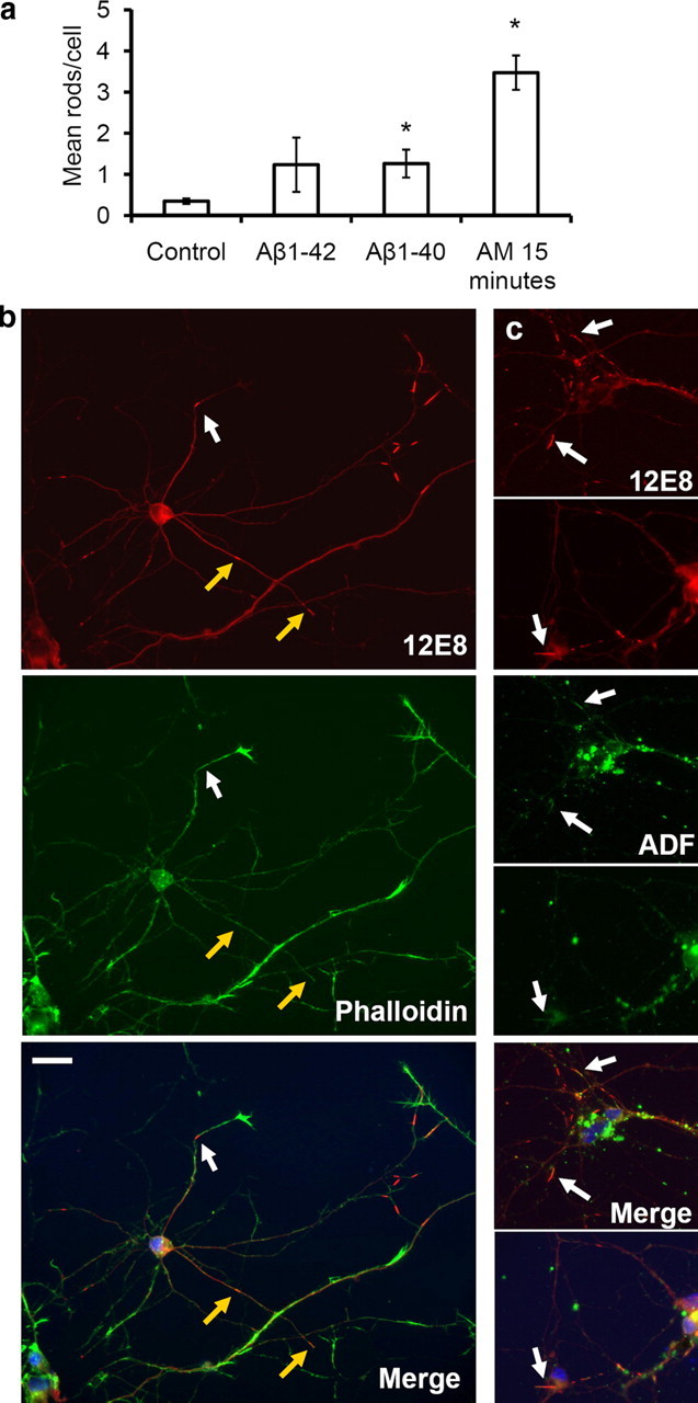
Amyloid peptides enhance pMAP sequestration to rods in primary chick neurons. Primary neurons were treated for 20 h with solutions of Aβ peptides (1 or 2 μm) and double-labeled with the 12E8 antibody (red) and either phalloidin-488 or ADF (both green) as indicated. a, The mean number of pMAP-positive rods per cell was quantified in >10 fields on control coverslips and coverslips treated with Aβ peptides for 20 h or AM for 15 min. pMAP-positive rods per cell increased significantly after incubation with AM or Aβ solutions compared with vehicle (DMSO) control (mean ± SEM, 1.3 ± 0.3 rods/nuclei for both aged Aβ1-40 and Aβ1-42 and 3.5 ± 0.4 rods/nuclei for AM-treated compared with 0.4 ± 0.1 rods/nuclei for control cells; *p < 0.02; n > 20; results are from duplicate experiments). Since no difference was seen between 1 versus 2 μm peptide treatments, results have been pooled. b, pMAP-positive rods in Aβ-treated neurons (shown here for Aβ1-40) often formed at distal regions near the base of phalloidin-positive (F-actin-rich) active growth cones (white arrows). They also formed in neurite shafts associated with collapsed growth cones (b, yellow arrows). c, Rods assembling in Aβ-treated neurons (shown here for Aβ1-40) accumulated both pMAP (red) and ADF (green) (arrows). Merged images (plus DAPI in blue) are shown in bottom panels. Scale bar, 20 μm.