
Figure 1.
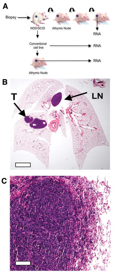
Generation and characterization of the SCLC primary xenograft model. A, Outline of the experimental approach from primary sample, xenograft, cell line and secondary xenograft from the derived cell line. B, H&E stained section of mouse lungs following orthotopic injection of the LX22 xenograft. The “primary” tumor (T) and metastasis in mediastinal lymph node (LN) are highlighted. Scale bar = 1mm C, H&E stained section of the intrapulmonary tumor shown in B. Scale bar = 20μm.