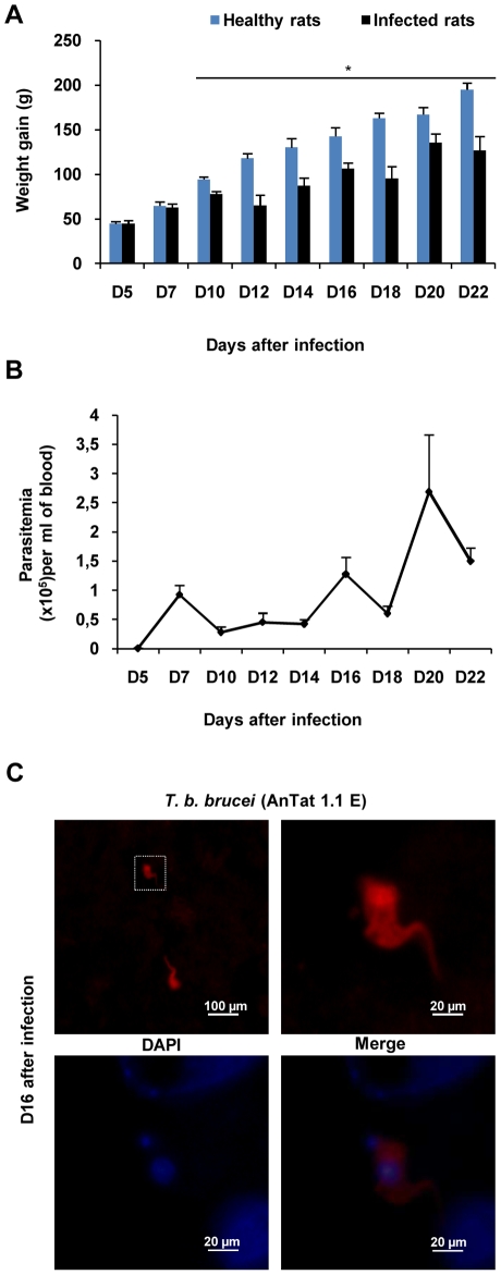
Figure 1. Biological diagnostic of experimental African trypanosomiasis in Wistar rats infected with T. b. brucei.
(A) Body weight gain in healthy and infected rats calculated from weight values at D0 before infection: mean value±SEM (* p<0.05 compared to healthy rats, n = 6 per day and per group). (B) Time course of parasitaemia, n = 6 per day). (C) Immunofluorescent staining of T. b. brucei (red) in the brain parenchyma at D16 post infection (n = 3). Nuclei were stained with DAPI (blue). (Microscope Olympus BX51equipped with Olympus DP50 camera). Statistics analysis, ANOVA followed by post hoc Fisher's test. Abbreviations: T. b. brucei, Trypanosoma brucei brucei; DAPI, 4′, 6′ Di Amidino-2-Phenylindole.

