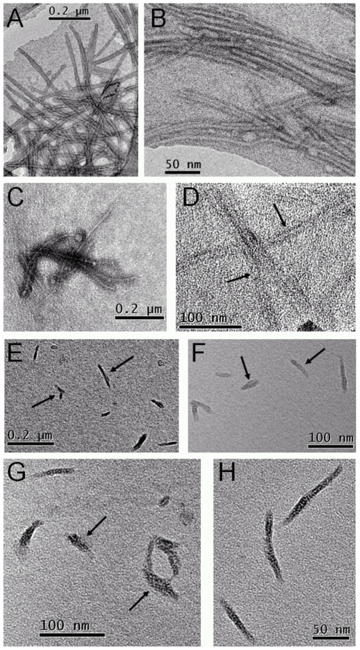
Figure 6. TEM micrographs showing the effect of the presence of 6-aminoindole or 5-hydroxindole on the aggregation of α-syn.
Whilst both samples revealed similar kinetic data, typical of a nucleation/polymerization process, microscopy revealed contrasting data. (A and B) α-syn fibrils formed in the absence of any ligands, (C and D) α-syn fibrils formed in the presence of 5-hydroxyindole and (E–H) 6-aminoindole. Arrows indicate fibrillar structures of varying size.

