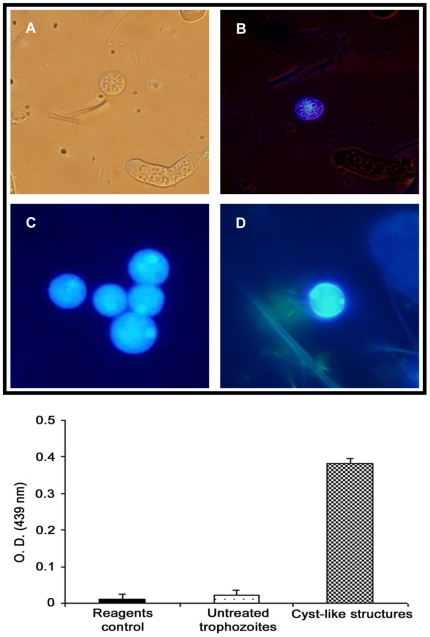
Figure 2. Detection of chitin in E. histolytica CLS resistant to detergents by calcofluor white staining (upper panel) and a WGA-binding assay (bottom graphic).
(A and B) Light and UV microscopy, respectively, of a mix culture of untreated trophozoites and CLS stained with calcofluor white. CLS is clearly differentiated from trophozoites by the spherical shape and small size. In B, blue fluorescence of CLS under UV contrasts with the absence of fluorescence in the trophozoites 20X; (C and D) Comparison of calcofluor staining of CLS (C) against an E. histolytica cyst isolated from human feces (D). In spite of both showing a similar blue-whitish fluorescence, staining of the surface is clearer in the cyst from human feces, whereas staining of some apparently internal structures (arrowheads), probably transporting vacuoles, is evident in the CLS (40X). In the bottom graphic, bars represent the standard deviations.

