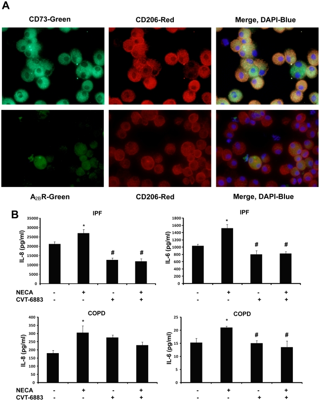
Figure 10. A2BR-dependent IL-8 and IL-6 expression in human primary alveolar macrophages.
(A) Cells from an IPF patient were reacted with antibodies against CD73 (A, upper panel, green) or the A2BR (A, lower panel, green) together with the M2 macrophage marker CD206 (red). In the merged images, yellow represents co-localization of CD73 or the A2BR and the M2 marker, blue is dapi stained nuclei. (B) ELISA measurements of IL-8 and IL-6 production from macrophage cultures of IPF and COPD patients. Results are presented as mean concentrations of cytokines ± SEM. *p≤0.05 versus cells without any treatment. #p≤0.05 versus cells treated with NECA alone. n = 6.

