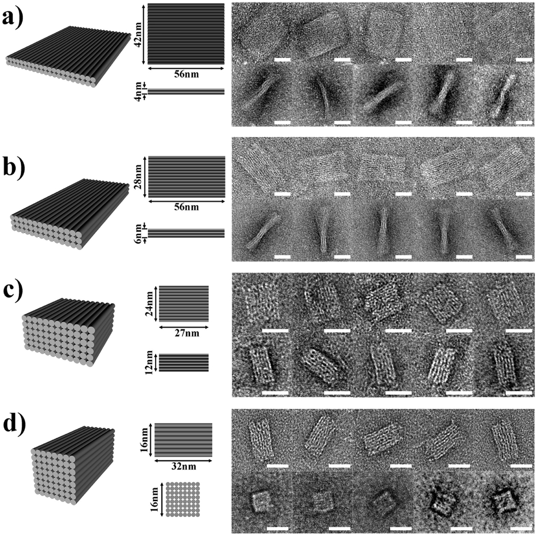
Figure 2.
3D DNA origami solid blocks. (a) Two-layer structure. (b) Three-layer structure. (c) Six-layer structure. (d) Eight-layer structure. The 3D perspective cylinder view and the projections of the top view and the side view are shown. Each cylinder represents a DNA double helix. For the 8-layer block in d, the end-view projection is shown. On the right are the representative transmission electron microscope (TEM) micrographs of negatively stained particles observed. The scale bars are 20 nm. For imaging, samples were adsorbed for 30 s onto glow-discharged grids (carbon-coated grid, 400 mesh, Ted Pella) and stained with 0.7% uranyl formate. Excess stain was wicked away by touching with a piece of filter paper, then dried at room temperature. The samples were imaged with a Philips CM200 microscope, operated at 200 kV in the bright field mode.

