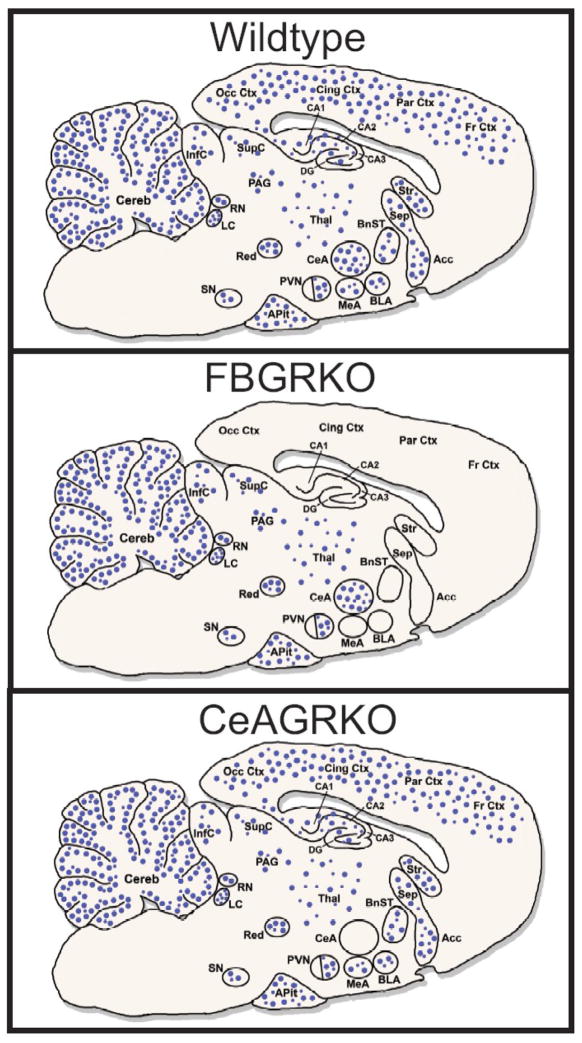
Fig. 1.
Expression of neuronal GR expression in wildtype, FBGRKO and CeAGRKO mice. GR is ubiquitously expressed throughout the brain, showing higher expression in a number of important limbic areas (e.g. CeA, PVN, hippocampus). Circles (●) represent neuronal glucocorticoid receptors (GR) in wildtype mice (top panel), FBGRKO mice (middle panel) and CeAGRKO mice (lower panel). Abundance of receptors is given by the relative density of circles in an area. Acc – nucleus accumbens; APit – anterior pituitary gland; BLA – basolateral nucleus of the amygdala; BnST – bed nucleus of the stria terminalis; CA1, CA2, CA3 – hippocampal areas CA1 to CA3; CeA – central nucleus of the amygdala; Cereb – cerebellum; Cing Ctx – cingulate cortex; DG – dentate gyrus; Fr Ctx – frontal cortex; InfC – inferior colliculus; LC – locus coeruleus; MeA – medial nucleus of the amygdala; Occ Ctx – occipital cortex; PAG – periaqueductal gray; Par Ctx – parietal cortex; PVN – paraventricular hypothalamic nucleus; Red – red nucleus; RN – raphe nuclei; Sep –septum; SupC – superior colliculus; SN – substantia nigra; Stri – striatum; Thal – thalamus.
Adapted from (Boyle et al., 2006; Kolber et al., 2008a; Kretz et al., 2001; Morimoto et al., 1996; Steckler and Holsboer, 1999).

