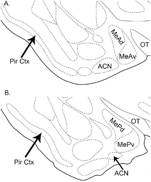
Figure 2.
Schematic of areas of interest in all 3 experiments. A. The anterior medial amygdala (MeA) is subdivided into dorsal (MeAd) and ventral (MeAv) portions. B. The posterior medial amygdala (MeP) is subdivided into dorsal (MePd) and ventral (MePv) portions. The anterior cortical nucleus (ACN) and piriform cortex (Pir Ctx) were counted in both sections and averaged (see Experimental Procedures). Modified from Morin and Wood (2001). MeA sections correspond to Fig. 26, AP −1.2 mm from bregma; and MeP sections correspond to Fig. 28; AP −1.8 mm from bregma in the Morin and Wood atlas.

