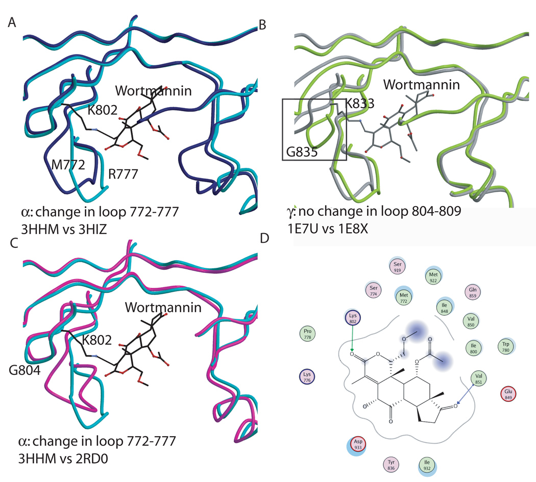
Figure 6. Wortmannin binding to the p110α and γ kinase domains.
A. Conformational changes elicited by wortmannin binding to p110α (H1047R p110α + wortmannin in turquoise; H1047Rp110α in blue). B. Conformational changes observed in p110γ in response to wortmannin binding. The black box highlights one of the observed differences in residues 748–750, 832–838, 871–876 which moved away from the wortmannin. C. Comparison between the changes due to wortmanninn binding in p110α (turquoise) and p110γ (grey). D. Interaction between wortmannin and the p110α structure.

