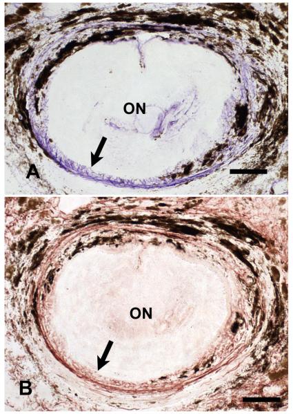
Figure 1.
Peripapillary sclera of young C57/BL6 mouse in cross-section. The upper micrograph shows a dense ring of purple, Luna-stained elastin fibers (arrow) is present immediately adjacent to the optic nerve head. A serial section from the same sample (bottom) that is stained with rabbit anti-elastin primary antibody demonstrates that the material stained by the Luna technique is similar in configuration to that identified by anti-elastin (arrow). (Top x20, bar = 40um; bottom x20, bar = 50um)

