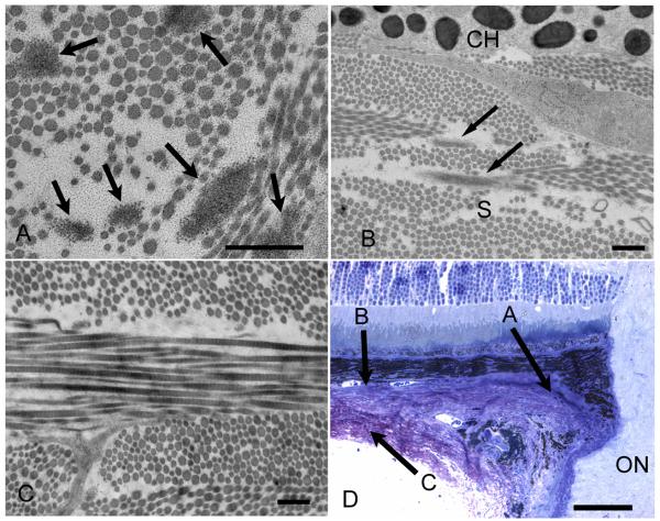
Figure 5.
Transmission electron micrograph of C57/BL6 mouse sclera in cross-section. A. Six elastin fibers (arrows) are present in the peripapillary sclera, the region where elastin is most dense (x12,000, bar=500nm). B. Elastin is present (arrows) in the innermost sclera adjacent to the choroid, whose melanocytes are seen at top (x10,000, bar=500nm). C. Outer sclera region showing no elastin present (x10,000, bar=500nm). D Light micrograph demonstrating the regions that each of the transmission electron micrographs is representing (x40, bar=50um). Variable diameter collagen fibrils are seen throughout the sclera in all images.

