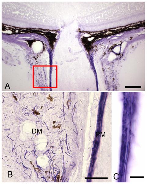
Figure 6.
The upper light micrograph (x20, bar=40 um) demonstrates the location of the magnified images (bottom) of Luna-stained elastin in the meninges. Elastin (arrows) is present in both the dura mater (DM) and pia mater (PM). B. Dura mater elastin shows no preferred orientation, with cross-sectioned fibers seen as dots or short segments and longitudinal fibers seen as extended lines (x63, bar = 30um). C. Pia mater elastin is arranged in two layers: an outer zone in which fibers run around the circumference of the nerve (seen here as dots) and an inner layer (seen as linear profiles) parallel to the long axis of the optic nerve (ON) (x100, bar = 10um).

