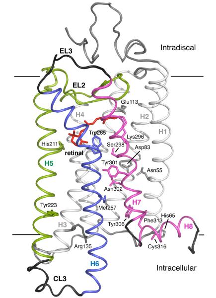
Fig. 1.
Crystal structure of rhodopsin. The photoreactive 11-cis retinal chromophore (red) is buried within the bundle of seven TM helices on the extracellular (or intradiscal) side of the receptor. Transmembrane helices H1-H4 (gray) form a rigid framework that is stabilized by tight packing mediated by group conserved amino acids and hydrogen bonding interactions.100 Guided MD simulations are used to characterize the motion of TM helices H5 (green), H6 (blue) and H7 (purple) upon isomerization of the retinal and deprotonation of the retinal – Lys296 Schiff base linkage.

