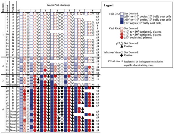
Fig. 3.
Outcome of FeLV challenge. Blood samples collected pre-challenge (0 weeks PC) and weekly thereafter were assessed for FeLV DNA (blue squares) and RNA (red circles) by qPCR, for p27 (triangles) by capture ELISA, and for infectious FeLV (diamonds) by VI at 4 weeks PC only. The VN antibody response was assessed post-vaccination pre-challenge and again at 8 weeks PC. The range of viral DNA and RNA levels is expressed by the intensity of the blue or red, respectively.

