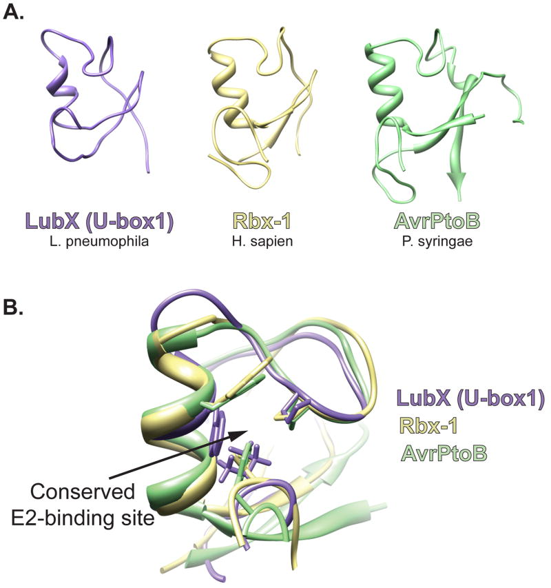
Figure 1.
Bacterial mimics of eukaryotic RING/U-box E3 ligases. (A). Using the Phyre threading program, the sequence of U-box1 of L. pneumophila LubX was aligned to known structures and the structure was modeled to its best fit, human E3 traf6 (E-value of 2.6e−11; estimated precision of 100%); the RING/U-box structure of H. sapien, Rbx-1 (PDB ID 3DPL); the core fold of P. syringae, AvrPtoB (PDB ID 2FD4). (B) Visualization of the E2-binding site residues of Rbx-1 with homologous regions in LubX and AvrPtoB. The three putative E2-binding residues are shown.

