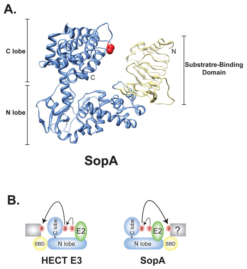
Figure 2.
SopA is a HECT-like E3 ligase. (A) Overall structure of SopA163–782 (PDB ID 2QYU); the HECT domain of SopA is shown in blue and the N-terminal β-helix domain in yellow. The catalytic cysteine is signified in red. N, NH2 terminus; C, COOH terminus. (B) Schematic diagram of the transfer of ubiquitin. Ubiquitin is shown in red. The C lobe of a generic HECT E3 or SopA is positioned similarly to accept ubiquitin following binding of Ub-charged E2 (green). However, the placement of the substrate binding (SBD; yellow) domain of SopA on the opposite end of the N lobe as found in eukaryotic HECT E3s likely requires a significantly different conformational change to facilitate ubiquitin transfer to a target substrate (grey box).

