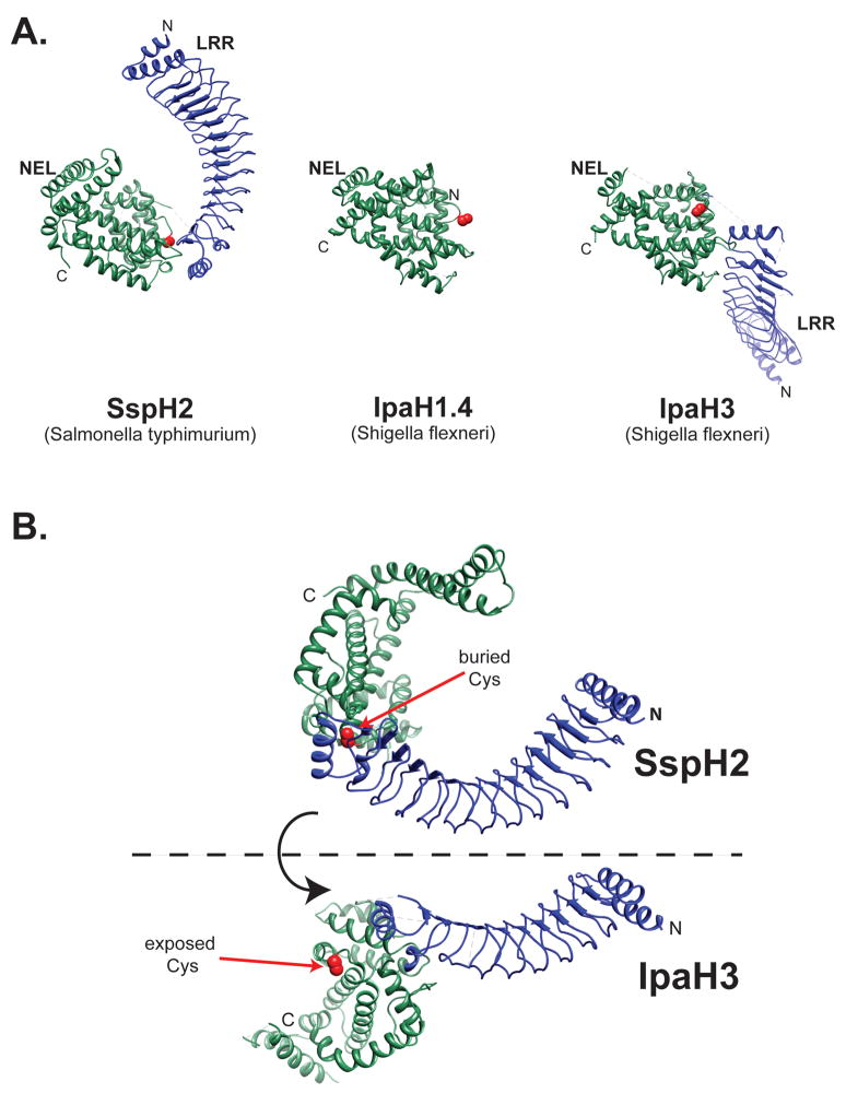
Figure 3.
NEL family of E3 ligases. (A) The structure of SspH2166–783 (PBD ID 3G06), IpaH1.4265–575 (PDB ID 3CKD), and Ipa325–561 (PDB ID 3CVR) is shown with leucine-rich repeat (LRR) domain in blue and the novel E3 ligase (NEL) domain in green. The catalytic cysteine residue is shown in red. N, NH2 terminus; C, COOH terminus. (B) Conformational changes likely activate the NEL family of E3 ligases. The structures of Shigella IpaH3 and Salmonella SspH2 suggests a dramatic hinge motion as the NEL domain rotates 180° from the closed conformation indicated by the structure of SspH2 to the open position represented by the IpaH3 structure. The catalytic cysteine residue is shown in red. N, NH2 terminus; C, COOH terminus.

