
Fig. 1.
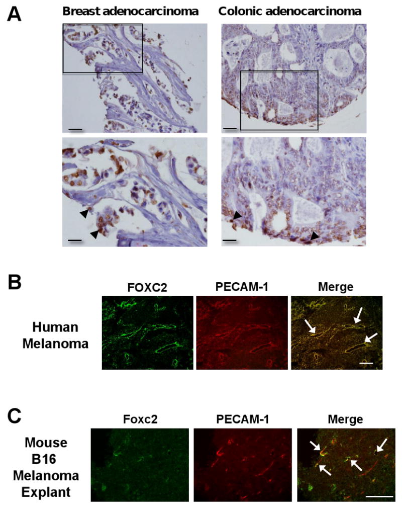
Expression of human and mouse FoxC2. (A) FOXC2 expression in the human breast and colon tumors. FOXC2 expression in lobular adenocarcinoma of breast and colonic adenocarcinoma at 20× (upper panels) and 40× (lower panels) magnifications; the areas presented at higher magnification are indicated by square. Note that FOXC2 immunostaining is primarily limited to cancer cells in tumor tissue. Scale bars, 500 μm and 100 μm for upper and lower, respectively. (B) Expression of FOXC2 is detected in tumor endothelial cells of human malignant melanoma. Frozen sections of malignant melanoma in the human skin were co-immunostained with anti-FOXC2 and anti-PECAM-1 antibodies. FOXC2 protein (green) was detected in tumor endothelial cells positive for PECAM-1 (red). Arrows indicate FOXC2-positive human endothelial cells. Scale bar, 250 μm. (C) Expression of Foxc2 is detected in endothelial cells of mouse B16 melanoma. Co-immunostaining with anti-FOXC2 and anti-PECAM-1 antibodies in mouse B16 tumors grown in C57BL/6 mice for 11 days after subcutaneous injection. Arrows indicate overlapping expression of Foxc2 (red) and PECAM-1 (green) in tumor endothelial cells. Scale bar, 250 μm.