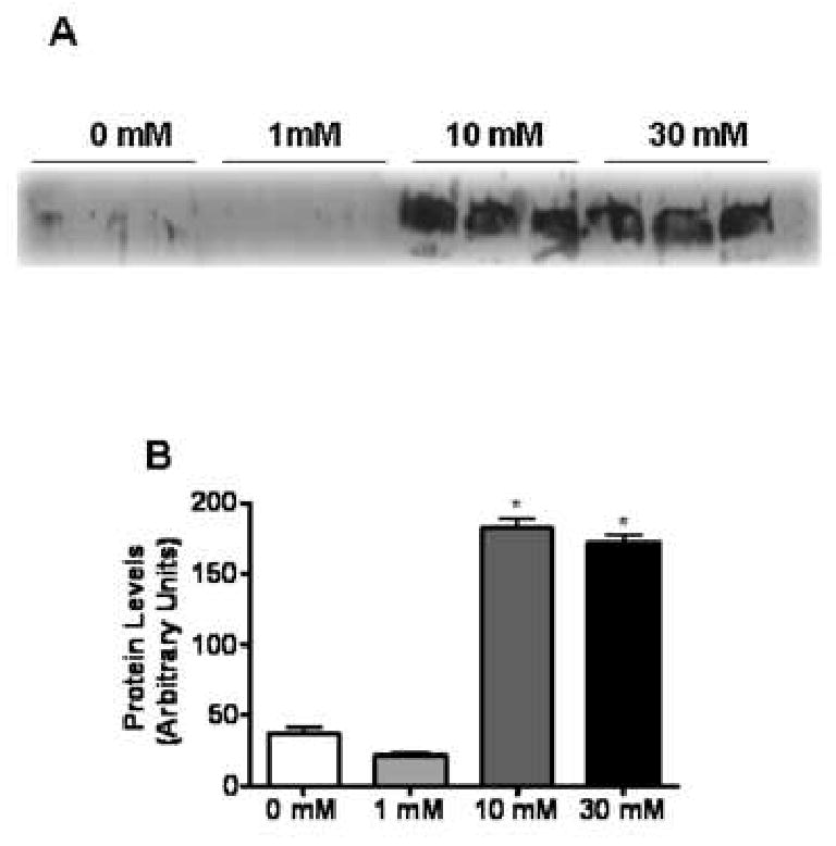
Fig. 8. Release of HMGB1 into the cellular supernatant of APAP-treated TAMH cells.

TAMH cells were treated with APAP (0, 1, 10 or 30 mM) for 15 hr. Following treatment, cellular supernatant was collected, concentrated and residual APAP was removed via a desalting column. (A) Immunoblot analysis of HMGB1 release into the cellular supernatant. (B) Quantification of HMGB1 release into the cellular supernatant by densitometry. *, P < 0.05, compared with cells treated with 0 mM APAP.
