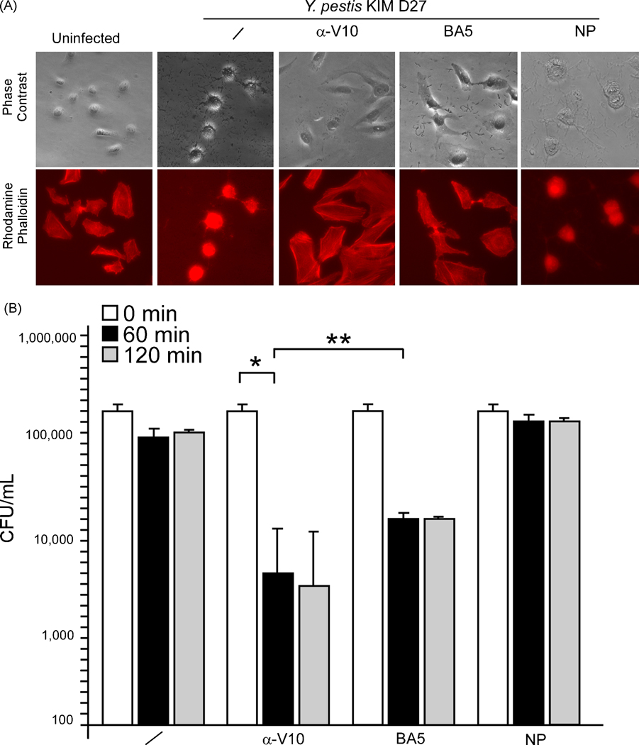
Fig. 2.
MAb BA5 blocks Y. pestis type III injection of host cells. (A) Y. pestis cytotoxicity for HeLa cells was measured by staining F-actin with Texas red-conjugated phalloidin. Differential interference contrast (DIC) images were captured at a magnification of 100×, and fluorescence was measured at 608 nm emission. Samples were compared with a well that received no bacteria (uninfected). At the time of HeLa cell infection, 100 µl of rV10 rabbit polyclonal sera (α-V10) or 500 µg of monoclonal BA5 (BA5) or 500 µg of non-protective LcrV monoclonal (NP) were added. (B) Human blood, anti-coagulated with lepirudin, was incubated for 60, 120 or 240 minutes with 1 × 10⁵ CFU Y. pestis KIM D27 in the absence (/) or presence of 100 µl of α-V10 rabbit polyclonal sera, 500 µg of monoclonal MAb BA5 or 500 µg of non-protective LcrV monoclonal antibody (NP). Bacterial load (CFU/ml) was recorded by plating aliquots on agar and incubating for colony formation. * P <0.001, ** P = 0.15.

