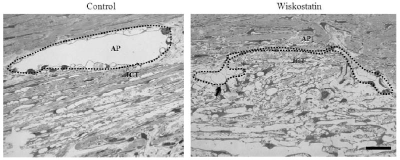
Figure 2.

Light microscope-based histological integrity of outflow tissues perfused with or without Wiskostatin. After 5 hr perfusion, 3 in 6 specimens from different quadrants of 2 eyes were found to have clear oval shaped aqueous plexi (AP) in the control group. In contrast, none in 6 specimens had a clear oval shaped aqueous plexi in Wiskostatin perfused group. Instead, some of them had deformed aqueous plexi (marked with a dotted line), into which JCT was found to be herniated and with prominent giant vacuoles (indicated with arrows) in the endothelial cells of aqueous plexi compared to controls. Scale bar: 20 μm.
