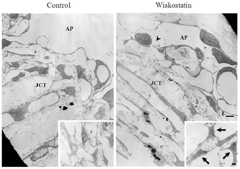
Figure 3.

Transmission electron micrographs of outflow tissue perfused with or without Wiskostatin. Drug perfused samples appear to have enlarged and more giant vacuoles in the inner wall of aqueous plexi compared to control specimens. A small break in the lining of endothelial cells (arrowhead), and enlarged TM cells (arrows) in the corneoscleral meshwork (insets) were found in a Wiskostatin perfused eye. Scale bar: 2 μm.
