
Figure 1.
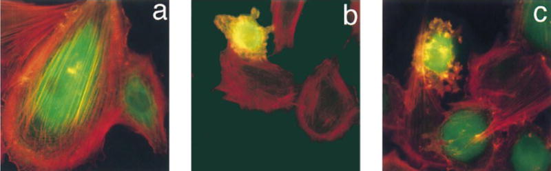
Changes in morphology and stress fiber formation in REFs stained with phalloidin for F-actin (Red) and FITC-IgG for microinjected cells (green). (A) REFs microinjected with caged PKA 4, (B) REFs microinjected with caged PKA 4 followed by photouncaging, (C) REFs microinjected with free PKA catalytic subunit. Reprinted with permission from [66]. Copyright 1998 American Chemical Society.