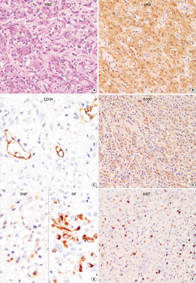
Fig. 3.
GANT reveals mainly epithelioid feature. Focal cytoplasmic vacuolization is seen. The tumor cells are robustly positive for CD117 (c-kit) but negative for CD34. S-100 protein, synaptophysin and NF are partially positive in neoplastic cells. Ki67 labelling index is high. (A: H&E, ×200, B: c-Kit, ×200, C: CD34, ×400, D: S100, ×200, E: left: Synaptophysin, right: Neurofilaments, ×400, F: Ki67 immunostaining, ×200).

