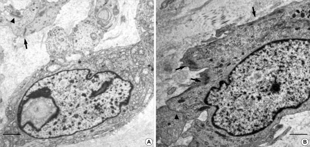
Fig. 6.
(A) Spindle cell GIST also shows cytoplasmic processes but do not have much villous processes. The neoplastic cells have a nuclear pseudoinclusion and rich organelles including electron dense lysosomal granules. There are hemidesmosome-like junctions (arrowheads) as well as a few gap junctions (arrow). Incontinuous external lamina is seen. (B) Ultrastructual differentiation toward smooth muscle, such as cytoplasmic myofilaments are seen (arrows) (uranyl acetate and lead citrate stain, bar: 2 µm, A: ×5,000, B: ×6,000).

