
Figure 4. Nile red staining visualizes deep tissue fat deposits in larval zebrafish.
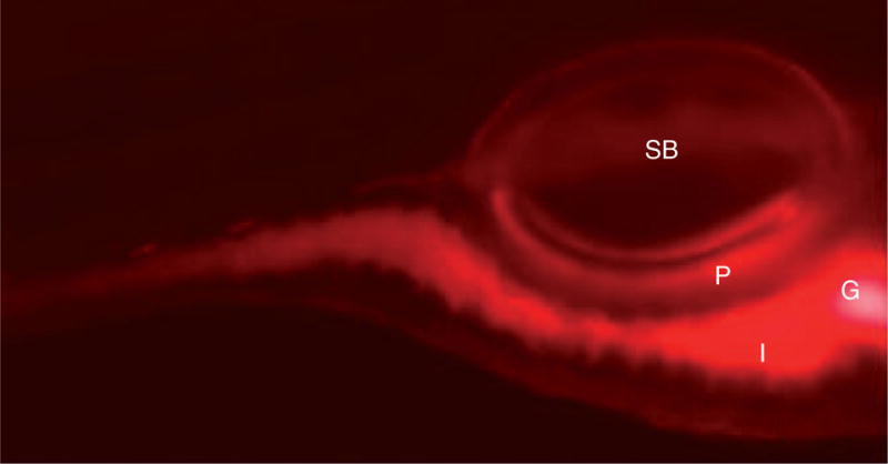
Extended incubation of larvae for at least 4 h in nile red (5 ng/ml) labels fat deposits. Nile red staining is present in the intestines (I), gall bladder (G) and pancreas (P). The swim bladder (SB) is also indicated.
Reprinted with permission from [87].