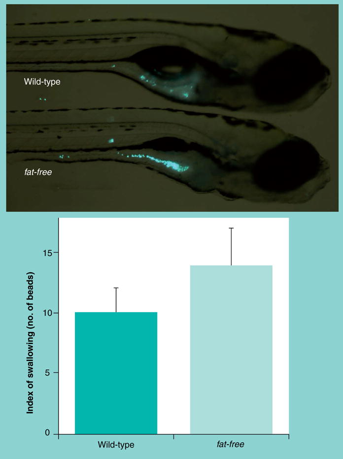
Figure 6. Microspheres reveal normal swallowing activity in the fat-free mutants.

Larvae (6 dpf) were placed in embryo media containing fluorescent latex microspheres (0.0025% Fluoresbrite plain YG 2.0um, Polysciences Inc.) for 1 h, washed, and imaged. Numbers of beads were 10 ± 2 in the wild-type versus 14 ± 3 beads in fat-free mutant larvae (mean ± SEM, n = 9; p > 0.3).
